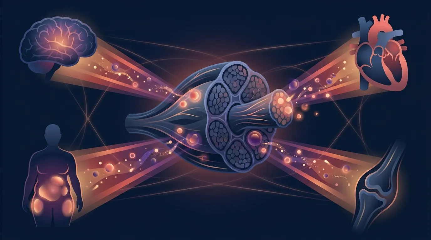
For most of the last century, skeletal muscle was viewed as a movement organ. You contracted it, it moved a limb, and that was the story. The metabolic and hormonal action was thought to happen elsewhere: pancreas, liver, thyroid, adipose tissue.
That view has changed.
The framing shift started in 2000, when Bente Klarlund Pedersen's group at the Centre for Inflammation and Metabolism in Copenhagen showed that contracting muscle releases interleukin-6 (IL-6) into the bloodstream in an exercise-dose-dependent way. By 2012, Pedersen and Mark Febbraio had articulated the modern framing in Nature Reviews Endocrinology: skeletal muscle is a secretory organ. The signaling molecules it releases are called myokines, and they act on distant tissue the same way a classical hormone does. This article walks through the primary evidence, the strongest randomized data, the mechanistic story, and what it actually means for how you should think about the movement you do.
The Research: What the Studies Actually Show
Pedersen and Febbraio 2012: The Framing Review
The single most-cited paper in this field is Pedersen and Febbraio (2012), "Muscles, exercise and obesity: skeletal muscle as a secretory organ," published in Nature Reviews Endocrinology. It is a review, not a randomized trial, but its purpose is to consolidate a decade of mechanism work into a coherent framing. The core claim: skeletal muscle expresses, produces, and releases myokines, and these myokines mediate many of exercise's systemic health benefits.
Three concrete pieces from the review that anchor the rest of the literature:
- IL-6 is the prototype myokine. During prolonged aerobic exercise, plasma IL-6 can rise up to 100-fold. Unlike chronic low-grade IL-6 elevation (which tracks with obesity and cardiovascular risk), acute muscle-derived IL-6 is transient, muscle-sourced, and metabolic in effect.
- Muscle-derived IL-6 has metabolic actions. It stimulates AMPK-mediated fat oxidation and hepatic glucose output during exercise, and it appears to have anti-inflammatory effects downstream by inducing IL-10 and IL-1 receptor antagonist.
- The class is larger than one molecule. By 2012, several dozen candidate myokines had been identified, and the review predicted the number would climb into the hundreds. It did.
Wedell-Neergaard 2019: The Causal RCT for IL-6 and Visceral Fat
Reviews propose mechanisms. Randomized trials test them. The best causal evidence connecting muscle-derived IL-6 to a real health outcome is Wedell-Neergaard et al. (2019) in Cell Metabolism. Fifty-three adults with abdominal obesity were randomized in a 2x2 factorial design to:
- 12 weeks of supervised aerobic exercise (cycling) or no exercise, and
- The IL-6 receptor blocker tocilizumab or placebo, infused every four weeks.
Visceral adipose tissue mass was measured by MRI at baseline and 12 weeks. The key finding:
- In the placebo arm, exercise reduced visceral fat significantly (about 8 percent).
- In the tocilizumab arm, exercise did not reduce visceral fat. In fact, visceral fat rose modestly in this arm.
- Cardiorespiratory fitness improved with exercise regardless of tocilizumab.
That double dissociation matters. Fitness gains are IL-6-independent. Visceral fat loss is IL-6-dependent. This is the strongest human evidence that a myokine mechanistically drives an exercise adaptation, not just correlates with it. It also flags something important. Long-term IL-6 blockade (used clinically for rheumatoid arthritis) may blunt one of exercise's specific metabolic effects. That is a real drug-exercise interaction, and it emerged from this trial.
Boström 2012: Irisin and Fat Browning
The most famous, and most contested, myokine story is irisin. Boström and colleagues (2012) in Nature showed that muscle-specific overexpression of PGC-1α (the master regulator of mitochondrial biogenesis) drove expression of FNDC5, a membrane protein that is cleaved and released into circulation as irisin. Irisin then acted on subcutaneous white adipose tissue to induce a "browning" program (UCP1 expression, brown-fat-like features), which in mice raised whole-body energy expenditure and improved glucose homeostasis.
Two things are worth knowing about the irisin literature.
First, the mouse mechanism has held up. PGC-1α drives FNDC5, FNDC5 is exercise-inducible, and there is a real muscle-to-adipose signaling axis. Second, the human quantitative claims from the original paper have been narrowed by later work. Several early commercial ELISA assays for circulating irisin turned out to be non-specific, which meant many mid-2010s "irisin correlates with X" papers used unreliable numbers. Careful mass spectrometry studies confirmed that irisin does circulate in humans, but at concentrations lower than the original assay-based reports suggested, and the browning-of-white-fat effect appears smaller in humans than in mice. Treat the mechanism as real. Treat the human effect size as uncertain.
Wrann 2013: FNDC5, BDNF, and the Muscle-Brain Axis
If you have ever seen the claim "exercise grows your brain," part of the mechanism runs through this paper. Wrann et al. (2013), "Exercise Induces Hippocampal BDNF through a PGC-1α/FNDC5 Pathway," in Cell Metabolism, showed that endurance exercise raises FNDC5 expression in the hippocampus itself, and that FNDC5 induction drives expression of brain-derived neurotrophic factor (BDNF), the neuronal growth factor most tightly linked to learning, memory, and mood.
The pathway they demonstrated:
- Endurance exercise raises PGC-1α in muscle (established) and in hippocampus (new finding).
- PGC-1α with the co-factor ERRα drives FNDC5 transcription.
- Overexpressing FNDC5 in cultured neurons raises BDNF expression. Knocking it down lowers BDNF.
- Delivering FNDC5 peripherally to mice via adenoviral vector raised circulating irisin and induced hippocampal BDNF and other neuroprotective genes.
This is one of several plausible mechanisms behind exercise's effects on memory and mood. It sits alongside direct hippocampal blood flow effects, the cortisol-BDNF story, and the muscle-derived cathepsin B story (a separate myokine, later shown to cross the blood-brain barrier and stimulate adult neurogenesis in the hippocampus). We covered the practical implications in our BDNF and exercise and exercise and anxiety research writeups.

Severinsen and Pedersen 2020: The Comprehensive Synthesis
By 2020, the field had grown enough to justify a comprehensive review. Severinsen and Pedersen (2020) in Endocrine Reviews catalogued hundreds of candidate myokines and organized their known effects by target organ. The review's contribution is not any single new experiment but rather a taxonomy: which myokines act on brain, on adipose, on bone, on liver, on gut, on vasculature, on skin, and on muscle itself. The scale is what surprises most readers. The muscle secretome is not a handful of signals. It is a dense communication system.
Chow 2022: Zooming Out to Exerkines
The most recent framing shift is the move from "myokines" (muscle-specific) to "exerkines" (any tissue, any exercise-triggered signal). Chow, Gerszten, Taylor and colleagues (2022) in Nature Reviews Endocrinology defined exerkines as signaling moieties released in response to acute or chronic exercise from muscle (myokines), heart (cardiokines), liver (hepatokines), white and brown adipose tissue (adipokines and baptokines), and neurons (neurokines). Their review makes the case that exercise's systemic effects are best understood as an inter-organ signaling event, not a muscle-only event. Lactate, historically a metabolic waste product, is one of the interesting examples. A 2022 Nature Reviews Endocrinology piece by others argued lactate should now be considered a major myokine and exerkine in its own right, with direct signaling roles that go far beyond acting as a fuel.
Why This Matters for Your Fitness
The reason this literature matters for how you train is not that you should chase any specific molecule. You cannot dose irisin at home. IL-6 is not a supplement you can take. The takeaway is systemic.
Every time you contract muscle at a meaningful intensity, you are running a signaling program that touches your brain, fat, liver, bone, and immune system. This is why the health effects of exercise scale so bizarrely across seemingly unrelated conditions. Depression, insulin resistance, sarcopenia, some cancers, dementia, cardiovascular disease. All of them share this feature: contracting muscle produces signals the disease process is sensitive to.
Three practical implications follow.
First, frequency of contraction matters as much as any single workout's intensity. A single long session releases a burst of myokines, and then things reset. Consistent contraction across the week produces a chronic signaling environment. This is one of the mechanistic reasons research on movement snacks (short bouts of activity spread across the day) shows metabolic and cognitive benefits out of proportion to the total time involved. We cover the epidemiology in our exercise snacks and cancer risk writeup.
Second, both aerobic and resistance work count. IL-6 spikes with prolonged aerobic exercise and with intense resistance work. Irisin/FNDC5 rises with endurance and resistance modes. Cathepsin B (the muscle-brain myokine) rises with running in humans. The Severinsen and Pedersen synthesis catalogues myokine responses across modes and does not single out one type of training as necessary. If you enjoy cycling, cycle. If you prefer strength training, lift. If you like both, do both. What you cannot do is stay sedentary and get the same signaling.
Third, the intensity does not need to be extreme. Brisk walking, resistance-band circuits, bodyweight strength sessions, yoga flows that involve real strength holds, and gentle cycling all engage muscle contraction hard enough to release myokines. Extreme intensity releases more, but you get a large fraction of the benefit at moderate intensities that most people can sustain long-term.
How Myokines Work in Practice
To make this concrete, consider what actually happens in your body over a single 30-minute workout.
You start warming up. Contraction begins, and within minutes IL-6 mRNA is upregulated in the working muscle. During the workout, IL-6 protein is released into the blood. If the session is prolonged enough, plasma IL-6 climbs sharply. Your liver ramps up glucose output in response. Fat oxidation in muscle rises. Downstream, IL-6 induces IL-10 and IL-1 receptor antagonist, both of which are anti-inflammatory. This is why the acute inflammatory-looking IL-6 spike from exercise is metabolically distinct from the chronic low-grade IL-6 elevation seen in obesity. Same molecule, different biology.
Meanwhile, FNDC5 expression is rising in the working muscle. Some FNDC5 is cleaved and released as irisin. It circulates. In the hippocampus, PGC-1α and ERRα drive FNDC5 expression there as well, which triggers BDNF. Muscle-derived cathepsin B enters circulation and crosses the blood-brain barrier, contributing separately to hippocampal signaling.
At the same time, muscle is releasing decorin (which suppresses myostatin, freeing muscle to grow), fractalkine (which appears to improve pancreatic beta-cell function), musclin (which acts on bone and heart), and BDNF itself (which acts locally as an autocrine signal on muscle growth). None of this is speculation. Each is documented in the Severinsen and Pedersen review and the Chow exerkines synthesis, with primary trial or preclinical citations.
You finish the workout. Most myokine levels return toward baseline within hours. But the downstream effects (transcription changes, receptor sensitivity shifts, tissue remodeling) unfold over days and weeks. When you exercise consistently, you are running this signaling program often enough that its cumulative downstream effects become the new set point.
Get an evidence-based plan built for you
FitCraft, our mobile fitness app, pairs you with an AI coach who builds you a personalized plan around your goals, schedule, and fitness level. Every FitCraft program is designed by Domenic Angelino, MPH (Brown University) and NSCA-CSCS, with research published in the Journal of Strength and Conditioning Research and Medicine & Science in Sports & Exercise.
Take the Free Assessment Free • 2 minutes • No credit cardCommon Misconceptions
Misconception 1: "Myokines Explain Everything About Why Exercise Works"
Not quite. Myokines are one important layer. There are others: direct mechanical loading effects on bone, tendon, and cartilage that require actual force through the tissue; direct blood flow effects on the brain; direct effects on autonomic tone; direct effects on the microbiome. The Chow exerkines review is careful about this. It argues that myokines and other exerkines are a big part of the mechanistic story, not the whole story. Treat "exercise works through myokines" as an insight, not a slogan.
Misconception 2: "You Need Special High-Intensity Training to Trigger Myokine Release"
Higher intensity does produce larger IL-6 responses in the acute setting. But myokines are released across a wide intensity range, and consistency is a stronger predictor of long-term outcome than peak intensity in any single session. The Wedell-Neergaard trial used cycling at moderate intensity (about 60 to 75 percent of maximum heart rate) for 12 weeks, and the visceral fat loss it produced was IL-6-mediated. You do not need to redline. You need to move regularly at an intensity that meaningfully contracts muscle.
Misconception 3: "Irisin Is a Weight Loss Hormone"
The marketing side of the wellness world has run ahead of the research here. In mice, irisin has striking effects on brown-fat-like adipose activation. In humans, the effect size on adipose browning appears to be smaller, the assays used in the mid-2010s wave of correlation papers were flawed, and irisin has not translated into any working weight-loss therapy. The mechanistic story is real. The "irisin is why exercise burns fat" simplification is not supported. Weight loss from exercise is a multi-signal, multi-organ, multi-week process. Irisin is a piece, not the engine.
What the Research Suggests Going Forward
The last decade of myokine research has done three things.
It has re-framed skeletal muscle as an active endocrine organ, not a passive movement effector. That framing has already changed how sarcopenia, muscle mass, and inter-organ metabolic disease are studied. The old view held that carrying more lean mass was mostly about basal metabolic rate. The new view holds that lean mass is also a signaling capacity. Losing 20 pounds of muscle in later life is losing 20 pounds of hormone-releasing tissue that was talking to your brain.
It has given us mechanistically satisfying stories for effects that used to be described only correlationally. Exercise reduces depression risk. Why? Part of the mechanism runs through PGC-1α to FNDC5 to hippocampal BDNF, plus muscle-derived cathepsin B, plus IL-6's downstream anti-inflammatory effects. Exercise improves insulin sensitivity. Part of the mechanism runs through IL-6 signaling on liver and adipose, plus lactate as an inter-organ substrate and signal, plus direct muscle GLUT4 remodeling. Exercise reduces visceral fat. Part of the mechanism, per Wedell-Neergaard, requires IL-6 signaling.
And it has surfaced practical friction points. Chronic anti-inflammatory drugs that block cytokine signaling (tocilizumab for rheumatoid arthritis, IL-1 blockers for certain conditions) may attenuate some myokine-mediated benefits of exercise. This is worth knowing if you or a family member is on such a therapy. It does not mean stopping the drug, and it does not mean stopping exercise. It means the interaction exists and is measurable.
What the field still lacks: reliable, standardized human assays for many myokines, direct-to-target therapeutics that would translate the mouse data into human treatment, and a full understanding of which combinations of exercise mode, intensity, and duration maximize which myokine responses in which populations. Those are open questions being worked on now. What you can do with the current knowledge is straightforward. Move often, in ways that require real muscle contraction. The signaling handles the rest.

How FitCraft Fits
Most fitness apps are built around a single mode (strength only, cardio only, yoga only) or a single intensity level. The myokine research argues the opposite. What matters is regular, varied muscle contraction across the week, not a single perfect protocol.
FitCraft pairs you with an AI coach who builds your plan around the goals, schedule, and fitness level you share at signup. The programs blend strength, cardio, mobility, and bodyweight work in a way that keeps you contracting muscle several times a week without needing you to decide the mix. For someone building the habit for the first time, that structure is what turns "I should move more" into a signaling program the research says your organs respond to.
If you want to see the broader research on how consistency (not intensity) drives real health outcomes, our consistency not intensity guide covers the epidemiology side. On the resistance-training-specific health outcome data, our resistance training and mortality writeup covers the large cohort evidence.
References
- Pedersen BK, Febbraio MA. "Muscles, exercise and obesity: skeletal muscle as a secretory organ." Nature Reviews Endocrinology. 2012;8(8):457-465. doi:10.1038/nrendo.2012.49 (PMID 22473333).
- Boström P, Wu J, Jedrychowski MP, et al. "A PGC1-α-dependent myokine that drives brown-fat-like development of white fat and thermogenesis." Nature. 2012;481(7382):463-468. doi:10.1038/nature10777 (PMID 22237023).
- Wrann CD, White JP, Salogiannnis J, et al. "Exercise Induces Hippocampal BDNF through a PGC-1α/FNDC5 Pathway." Cell Metabolism. 2013;18(5):649-659. doi:10.1016/j.cmet.2013.09.008 (PMID 24120943).
- Wedell-Neergaard AS, Lehrskov LL, Christensen RH, et al. "Exercise-Induced Changes in Visceral Adipose Tissue Mass Are Regulated by IL-6 Signaling: A Randomized Controlled Trial." Cell Metabolism. 2019;29(4):844-855.e3. doi:10.1016/j.cmet.2018.12.007 (PMID 30595477).
- Severinsen MCK, Pedersen BK. "Muscle-Organ Crosstalk: The Emerging Roles of Myokines." Endocrine Reviews. 2020;41(4):594-609. doi:10.1210/endrev/bnaa016 (PMID 32393961).
- Chow LS, Gerszten RE, Taylor JM, et al. "Exerkines in health, resilience and disease." Nature Reviews Endocrinology. 2022;18(5):273-289. doi:10.1038/s41574-022-00641-2 (PMID 35304603).
Frequently Asked Questions
What are myokines?
Myokines are signaling molecules (mostly peptides and small proteins) that skeletal muscle produces and releases into circulation, mainly in response to muscle contraction. Pedersen and Febbraio proposed the framing that skeletal muscle is a secretory or endocrine organ in their 2012 Nature Reviews Endocrinology review. The Severinsen and Pedersen 2020 Endocrine Reviews review estimates muscle can produce and secrete hundreds of these signaling factors, which act on muscle itself (autocrine), on nearby tissue (paracrine), or on distant organs like brain, fat, bone, and liver (endocrine). IL-6 is the prototype: it was the first molecule shown to be released from contracting muscle in a meaningful, exercise-dose-dependent way.
What is the difference between myokines and exerkines?
Myokines are the subset that come from skeletal muscle. Exerkines is the broader umbrella term. Chow and colleagues (2022, Nature Reviews Endocrinology) defined exerkines as signaling molecules released in response to acute or chronic exercise from any tissue, including muscle, liver, adipose tissue, heart, and gut. Every myokine is an exerkine, but not every exerkine is a myokine. The umbrella term matters because exercise adaptations turn out to involve inter-organ signaling, not just muscle-secreted factors.
Is irisin real?
The core biology is real; some of the early human claims have been narrowed by later research. Boström and colleagues discovered irisin in 2012 (Nature), showing that muscle contraction produces a cleaved fragment of FNDC5 that browns white adipose tissue in mice. In humans, the effect size on adipose browning appears to be smaller than the initial paper implied, and early commercial ELISA assays were later shown to be non-specific, which called some human circulating-irisin numbers into question. What has held up: FNDC5 is exercise-induced, the PGC-1α/FNDC5 pathway drives hippocampal BDNF expression (Wrann et al., 2013), and irisin remains an active target in metabolic research. Treat human effect sizes with more caution than mouse data.
Does IL-6 from muscle actually help you lose visceral fat?
The best causal evidence in humans is the Wedell-Neergaard 2019 randomized controlled trial in Cell Metabolism. Fifty-three adults with abdominal obesity were randomized in a 2x2 design to 12 weeks of exercise (cycling) or no exercise, and to the IL-6 receptor blocker tocilizumab or placebo, given every four weeks. In the placebo arm, exercise reduced visceral adipose tissue mass significantly. In the tocilizumab arm, that reduction was blocked. Cardiorespiratory fitness improved with exercise in both arms regardless of tocilizumab (4.44 mL/min/kg in the placebo arm and 3.11 mL/min/kg in the tocilizumab arm, both significant). That double-dissociation is why IL-6 is now considered a mechanistic driver of exercise-induced visceral fat loss, not just a passenger marker.
Do you need to lift heavy to get the myokine benefits of exercise?
No. Contraction is what releases myokines, and both aerobic and resistance exercise produce robust myokine responses. IL-6 spikes strongly with prolonged aerobic exercise (the Pedersen work) and with intense resistance work. Irisin/FNDC5 rises with both endurance and resistance exercise. The Severinsen and Pedersen 2020 review synthesizes myokine responses across exercise modes and concludes the useful frame is that contracting muscle is the release trigger, not any single sport or load. Bodyweight training, resistance-band work, cycling, walking briskly enough to raise heart rate, and dedicated strength sessions all count.